How do you prove hair regrowth in a clinical study?

Quick Answer: How is hair regrowth proven in a clinical study?
- Hair regrowth is proven with objective measurements taken at baseline and repeated at fixed time points, not with "before and after" impressions alone.
- The strongest studies combine standardized clinical photography, expert grading, instrument-based measurements and participant self-assessment, so each result confirms the others.
- Common instrumental tests include digital image analysis, phototrichograms, standardized hair-shedding (wash) tests, scalp barrier (TEWL) measurements and electron microscopy of the hair shaft.
- MDhair's 24-week, IRB-approved, third-party clinical study, published in the Journal of Drugs in Dermatology in 2025, used five layers of evidence and found significant improvements in hair growth, coverage and thickness (P<0.001) [1].
- In that study, shedding fell by 37.3% at 12 weeks, scalp water loss fell by 69% at 24 weeks, and 88.9% of participants reported overall hair improvement, with no adverse events [1].
Why is hair regrowth so hard to measure?
This section explains why a mirror and a phone camera are not enough to prove that hair is growing.
Hair regrowth is hard to measure because hair changes slowly, varies from day to day, and looks different under every light, angle and hairstyle. A single scalp hair grows only about 1 cm a month, and each follicle cycles through growth (anagen), transition (catagen) and rest (telogen) on its own schedule [6]. That means real improvement builds gradually over three to six months, while everyday factors like washing frequency, styling products and lighting can make hair look thicker or thinner overnight.
Clinical researchers solve this problem by standardizing everything: the same camera, lighting, head position and hair preparation at every visit, the same washing protocol before shedding counts, and the same room temperature and humidity before scalp measurements. When conditions are held constant, the only thing left to change is the hair itself.
Good studies also measure hair from several independent angles. Photographs show what people see, instruments capture what the eye cannot, and questionnaires record what participants feel. When all three point in the same direction, the result is far more convincing than any single measurement on its own [6].
What are the main methods used to measure hair growth?
Dermatologists group hair evaluation methods into non-invasive, semi-invasive and invasive techniques.
Most modern hair studies rely on non-invasive methods, which can be repeated at every visit without discomfort. A comprehensive review in the International Journal of Trichology divides the main techniques into the following categories [6]:
- Global photography: standardized images of the top, front and back of the scalp, used for expert grading and visual comparison over time [2].
- Phototrichogram and TrichoScan: a small area of scalp is clipped and photographed through a dermatoscope, and software counts hairs, measures their thickness and estimates the ratio of growing to resting hairs [3].
- Hair counts and shedding tests: hairs lost during a standardized wash or 60-second combing are collected and counted to track shedding [4].
- Hair mass and diameter: tools such as cross-section trichometry measure the bulk of a bundle of hair, reflecting both density and thickness [7].
- Scalp biophysics: devices such as the Tewameter measure transepidermal water loss (TEWL), a sign of scalp barrier health [10].
- Microscopy of the hair shaft: scanning electron microscopy (SEM) reveals the condition of the cuticle, the protective outer layer of each strand [9].
- Investigator and subject assessments: structured rating scales completed by trained experts and by participants themselves.
Semi-invasive methods such as the trichogram (plucking about 50 hairs to examine their roots) and invasive methods such as scalp biopsy are used mainly in diagnosis or specialized research. They are rarely needed to show cosmetic improvement over time [6].
How did MDhair's JDD clinical study measure hair regrowth?
This section expands on every clinical and instrumental test used in the MDhair trial.
MDhair's study, titled "Artificial Intelligence-Based Personalization of Treatment Regimen for Hair Loss: A 6-Month Clinical Trial," was published in the Journal of Drugs in Dermatology in March 2025 [1]. It was an IRB-approved study run by an independent, third-party clinical research center in the United States. The trial enrolled 38 women aged 34 to 65 with self-reported hair thinning, and 27 completed the full 24 weeks.
Each participant uploaded scalp photos and answered a questionnaire through MDhair's web app. MDhair's AI model, trained on about 47,000 images of people with hair loss, analyzed the photos and built a personalized kit combining topical serums, shampoos, oral supplements and marine collagen peptides. Participants were assessed at baseline and at follow-up visits through week 24, with the main objective measurements taken at baseline, week 12 and week 24.
The study measured results in five complementary ways.
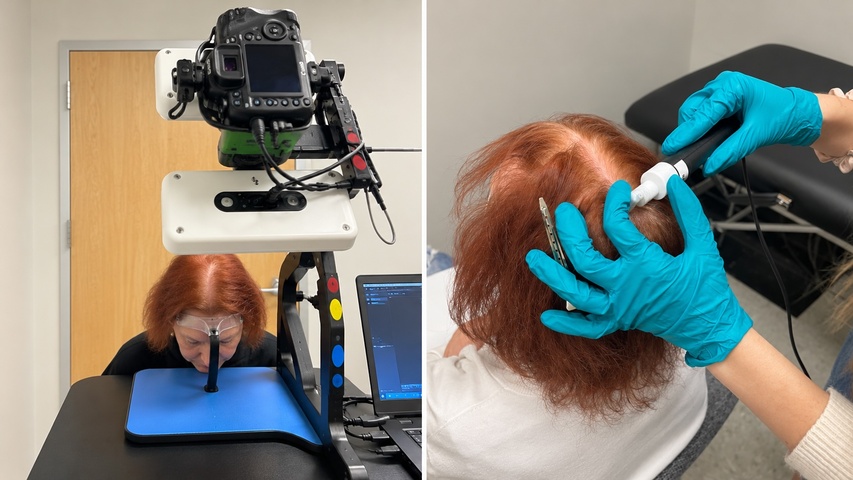
1. Standardized clinical photography (Canfield Hair Rig Imaging System)
All photographs were taken with the Canfield Hair Rig Imaging System, a professional imaging setup widely used in dermatology trials. It locks the head into the same position at every visit and uses fixed camera distance, angle and lighting, so that differences between images reflect real changes in the hair rather than changes in how the photo was taken [2].
This matters because even small shifts in lighting or head tilt can make thinning areas look larger or smaller. Standardized photography is the foundation that makes every other image-based measurement in the study reliable.
2. Expert clinical grading on a 7-point scale
Trained expert graders compared each participant's baseline and follow-up photographs and rated the change in hair growth, hair coverage, volume, thickness and overall fullness on a 7-point Likert scale, ranging from marked worsening through no change to marked improvement.
Expert grading of standardized photographs is the same approach used in pivotal hair-loss drug trials, where investigator assessments are a key outcome alongside hair counts [8]. In the MDhair study, expert grading showed statistically significant improvements in hair growth, coverage and thickness at both week 12 and week 24 (P<0.001), and about four in five participants showed improved hair growth by week 24 [1].
3. AI digital image analysis of scalp coverage
To add an objective, computer-based measure, the scalp photographs were also analyzed with digital image analysis. The software maps visible bare scalp in each image, highlighting thinning areas in white, so the area of exposed scalp can be compared across visits.
A smaller exposed area at follow-up means more of the scalp is covered by hair. Because the same algorithm is applied to every image, this method removes the human element entirely and provides a consistent, reproducible measure of hair coverage.
4. Standardized hair-shedding wash test
Shedding was measured with a standardized wash test. Participants washed their hair under a set protocol, and all hairs lost during the wash were collected and counted. Wash tests like this are a recognized way to quantify shedding and are based on the modified wash test described by Rebora and colleagues [4].
Shedding is often the first thing to change when hair health improves, well before new growth becomes visible. In the MDhair study, the average number of shed hairs fell by 37.3% at 12 weeks and 32.4% at 24 weeks, and most participants had lower shed counts at both time points (P<0.05) [1].
5. Scalp barrier function: transepidermal water loss (Tewameter)
Scalp health was measured with a Tewameter, a probe that measures transepidermal water loss (TEWL), the amount of water evaporating through the skin. A healthy skin barrier holds water in, so lower TEWL values indicate a stronger, better-functioning barrier [10]. Measurements are taken after participants rest in a temperature- and humidity-controlled room, which keeps readings consistent between visits.
A healthy scalp barrier supports a calmer, less inflamed environment around the follicles. In the MDhair study, scalp TEWL fell by 61.5% at 12 weeks and 69% at 24 weeks, showing a substantial improvement in scalp barrier function [1].
6. Scanning electron microscopy (SEM) of the hair shaft
To look at the hair itself at very high magnification, hair samples were examined with a JEOL JSM-6490LV scanning electron microscope at the North Dakota State University Electron Microscopy Core. SEM shows the overlapping scales of the cuticle, the outer layer that protects each strand and gives hair its shine and strength.
Cuticle condition is commonly scored on a standard grading system, from Grade 0 (healthy, intact, regularly overlapping cuticle) to higher grades showing lifted scales, cracks, holes and eventually exposed inner cortex [9]. In the MDhair study, SEM images showed improved hair texture, with representative strands moving from Grade 2 damage (lifted cuticle with cracks or holes) at baseline to a Grade 0, intact cuticle after six months of treatment [1].
7. Participant self-assessment questionnaires
Finally, participants rated their own results with a structured questionnaire covering nine aspects of hair and scalp health, including fullness, thickness, density, shedding, brittleness, frizz, shine and scalp health. Self-assessment captures what matters most to the people using the treatment: how their hair looks and feels in daily life.
In the study, 88.9% of participants reported overall hair improvement, 85.2% reported better scalp health and 92.6% reported less hair brittleness, all statistically significant (P<0.001) [1]. No adverse events were reported during the 24 weeks.
Why does combining several tests make results more convincing?
Each method answers a different question about the hair and scalp.
No single test captures everything that happens when hair improves. Photography and expert grading show visible growth and coverage. Image analysis confirms coverage changes with an objective algorithm. The wash test shows whether follicles are holding on to their hairs longer. TEWL shows whether the scalp environment is healthier, and SEM shows whether the hair fiber itself is stronger. Self-assessment confirms that these changes are noticeable in real life.
When measurements from independent methods agree, the overall finding becomes much more robust. In the MDhair study, every layer of evidence pointed in the same direction: more growth and coverage, less shedding, a stronger scalp barrier, healthier strands and high participant satisfaction [1].
| Test used in the MDhair study | What it measures | Key result |
|---|---|---|
| Canfield Hair Rig photography + expert 7-point grading | Visible hair growth, coverage, thickness, volume, fullness | Significant improvement at weeks 12 and 24 (P<0.001) |
| AI digital image analysis | Area of visible scalp (coverage) | Improved hair coverage over 24 weeks |
| Standardized wash test | Number of hairs shed per wash | 37.3% less shedding at 12 weeks; 32.4% at 24 weeks |
| Tewameter (TEWL) | Scalp barrier function | 61.5% lower at 12 weeks; 69% lower at 24 weeks |
| Scanning electron microscopy | Cuticle integrity of the hair shaft | Improved hair texture and cuticle condition |
| Self-assessment questionnaire | Participant-perceived results | 88.9% overall improvement; 92.6% less brittleness |
What makes a hair-growth study trustworthy?
These are the features to look for when you read any hair product's clinical claims.
- Ethics approval: an Institutional Review Board (IRB) reviews the protocol to protect participants and ensure the study is conducted properly.
- Independent testing: a third-party clinical research center runs the study and collects the data, rather than the brand itself.
- Enough time: because of the hair cycle, studies should run at least 12 weeks and ideally 24 weeks or longer.
- Objective instruments: results should include instrument-based measurements, not only questionnaires.
- Statistics: results should be reported with p-values showing that changes are unlikely to be due to chance.
- Peer-reviewed publication: publication in an indexed medical journal means the methods and results were reviewed by independent experts.
MDhair's study meets each of these criteria: it was IRB-approved, run by an independent research center, lasted 24 weeks, used multiple validated instruments, reported statistically significant results, and was published in the peer-reviewed Journal of Drugs in Dermatology [1].
What does this mean for your hair?
The study's design offers a practical roadmap for tracking your own progress.
You can borrow the logic of a clinical trial at home. Take monthly photos in the same spot, with the same lighting, the same part and dry, unstyled hair. Note roughly how much hair you see in the shower drain or on your brush on wash days. Give any regimen at least three months before judging it, and six months to see its full effect.
MDhair makes this easier by analyzing your scalp photos with the same AI technology tested in the study and adjusting your kit as your hair changes. Your personalized kit may include Regrowth or Restore Serum, Regrowth or Restore Shampoo, Regrowth or Restore Supplements, Marine Collagen and, when appropriate, Minoxidil 2% or 5%, combining DHT-balancing botanicals such as saw palmetto, scalp stimulators such as caffeine, antioxidants such as green tea extract, and peptides that support the follicle and the hair fiber.
Key Takeaways
- Hair regrowth is proven by standardized, repeated, objective measurements, not single before-and-after photos.
- The best studies combine photography, expert grading, instruments and self-assessment.
- MDhair's JDD study used Canfield photography, 7-point expert grading, AI image analysis, wash tests, Tewameter TEWL, SEM and questionnaires.
- Results included significant gains in growth, coverage and thickness (P<0.001), 37.3% less shedding and 69% lower scalp water loss.
- Look for IRB approval, independent testing, at least 12–24 weeks and peer-reviewed publication when judging any hair product's claims.
Frequently Asked Questions
How long does a hair growth clinical trial need to be?
At least 12 weeks, and ideally 24 weeks or more. Hair grows about 1 cm a month and follicles need time to move from the resting phase back into active growth, so shorter studies can miss real improvements.
What is a hair wash test?
A wash test counts the hairs that fall out during a standardized shampoo. Participants follow the same washing routine before each count, and all shed hairs are collected and counted. A lower count over time shows reduced shedding.
What does TEWL tell you about the scalp?
Transepidermal water loss (TEWL) measures how much water evaporates through the skin. Lower TEWL means the skin barrier is holding moisture better, which reflects a healthier scalp environment for hair follicles.
Why use an electron microscope to study hair?
A scanning electron microscope magnifies the hair surface thousands of times, revealing the condition of the cuticle scales. It shows whether strands are smoother and better protected, which relates to shine, strength and resistance to breakage.
What is the Canfield Hair Rig?
The Canfield Hair Rig is a professional clinical imaging system that holds the head in a fixed position with controlled camera distance, angle and lighting. It makes photographs taken weeks or months apart directly comparable.
Who conducted the MDhair clinical study?
The study was conducted by an independent, third-party clinical research center in the United States under IRB approval. It was published as Bhardwaj V, Rodgers N, Harth O, Harth Y in the Journal of Drugs in Dermatology, 2025;24(3):233–238.
Were there side effects in the MDhair study?
No adverse events were reported during the 24-week study.
How can I track my own hair regrowth at home?
Take photos once a month in the same location and lighting, with the same part and dry hair. Track shedding on wash days, and judge results after at least three months of consistent use.
References
- Bhardwaj V, Rodgers N, Harth O, Harth Y. Artificial intelligence-based personalization of treatment regimen for hair loss: a 6-month clinical trial. J Drugs Dermatol. 2025;24(3):233–238. doi:10.36849/JDD.8611
- Canfield D. Photographic documentation of hair growth in androgenetic alopecia. Dermatol Clin. 1996;14(4):713–721.
- Hoffmann R. TrichoScan: combining epiluminescence microscopy with digital image analysis for the measurement of hair growth in vivo. Eur J Dermatol. 2001;11(4):362–368.
- Rebora A, Guarrera M, Baldari M, Vecchio F. Distinguishing androgenetic alopecia from chronic telogen effluvium when associated in the same patient: a simple noninvasive method. Arch Dermatol. 2005;141(10):1243–1245.
- Olsen EA, Messenger AG, Shapiro J, et al. Evaluation and treatment of male and female pattern hair loss. J Am Acad Dermatol. 2005;52(2):301–311.
- Dhurat R, Saraogi P. Hair evaluation methods: merits and demerits. Int J Trichology. 2009;1(2):108–119.
- Wikramanayake TC, Mauro LM, Tabas IA, et al. Cross-section trichometry: a clinical tool for assessing the progression and treatment response of alopecia. Int J Trichology. 2012;4(4):259–264.
- Olsen EA, Dunlap FE, Funicella T, et al. A randomized clinical trial of 5% topical minoxidil versus 2% topical minoxidil and placebo in the treatment of androgenetic alopecia in men. J Am Acad Dermatol. 2002;47(3):377–385.
- Kim YD, Jeon SY, Ji JH, Lee WS. Development of a classification system for extrinsic hair damage: standard grading of electron microscopic findings of damaged hairs. Am J Dermatopathol. 2010;32(5):432–438.
- Fluhr JW, Feingold KR, Elias PM. Transepidermal water loss reflects permeability barrier status: validation in human and rodent in vivo and ex vivo models. Exp Dermatol. 2006;15(7):483–492.
Related Search Terms
how to prove hair regrowth, hair growth clinical trial methods, how is hair growth measured, hair wash test shedding count, TEWL scalp barrier, Tewameter scalp, scanning electron microscopy hair cuticle, Canfield hair imaging, expert grading hair photos, phototrichogram TrichoScan, MDhair clinical study, MDhair JDD study results, AI personalized hair loss treatment study, clinically proven hair growth products, IRB approved hair growth study
Find the most effective hair growth treatment for you by taking the free hair assessment.
Recommended Read
- Hair Treatments: Breakthrough Clinical Study: AI-Powered Hair Loss Solution by MDhair
- Ingredients: The Science of Hair Growth: How Ingredients Actually Work
- Hair Treatments: Build your hair growth stack: the science behind combining topical + oral treatments
- Hair Treatments: Hair shedding vs. hair loss?
Find the most effective hair growth products for you by taking the free hair assessment.
Enjoyed this article? Add MDhair as a preferred source on Google.